A 50 year old Hispanic male with known HTN, IDDM with bilateral diabetic retinopathy, and CAD presented to the ED after awakening with complete loss of vision in the right eye. He stated that prior to going to bed the night before, his vision was "normal" (for him) in the right eye. He denied any recent trauma, history of glaucoma, foreign body in the eye, eye pain, or new changes to his contralateral vision. He had suffered chronic vision impairment in both eyes, and undergone multiple laser surgeries in bilateral eyes for diabetic retinopathy. He was accompanied by his wife.
His eye examination revealed the following:
OS: 20/100 (baseline), OD: able to percieve hand motion only.
Lids/lashes: appeared grossly normal.
Sclera: without injection or foreign body.
Cornea: no gross abnormalities and negative florescin test OU.
Pupils: no hyphema/hypopion OU.
Direct light reflex: sluggish OD, normal OS.
Consensual reflex sluggish OD (i.e. when shining light into the right eye the left constricted sluggishly, but when shining light into the left eye, the right eye constricted normally.)
Remainder of slit lamp examination was unremarkable.
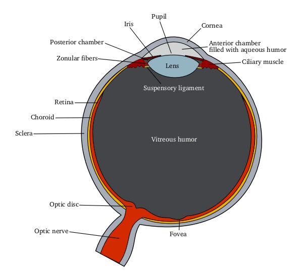
The Eye Schematic
From Wikipedia.com
Ophthomology consultation was ordered and the patient was evaluated.
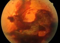
Fundoscopic evaluation revealed the following:
Secondary to suspicion for a particular diagnosis, ultrasound was performed and revealed the solution to the case.